Study of Appendicular Skeleton
Limb Bones
In tetrapod forelimb and hind limbs are present for locomotion and to support the body above ground level.
The overall patterns of the fore and hind limbs are similar. it is suitable to mention their general structure collectively. Thus, each fore and hind limb has the following segments.
1. Girdles
2. Stylopodium
3. Zygopodium
4. Autopodium

Table showing various segments of the fore limb and hind limb.
Segment
|
Forelimb
|
Hind limb
|
Stylopodium / Propodium
|
Humerus
|
Femur
|
Zeugopodium/ Epipodium
|
Radius, Ulna
|
Tibia, Fibula
|
Manus
|
Pes
| |
Autopodium/ Mesopodium
|
Carpals
|
Tarsals
|
Proximal row
|
a. Radial
b. Intermedium
c. Ulnar
|
a. Tibial
b. Intermedium
c. Fibular
|
Middle row
|
Central
|
Central
|
Distal row
|
Distal carpals
|
Distal tarsals
|
Metapodium
|
Metacarpals
|
Metatarsals
|
Phalanges
|
Phalanges
|
Phalanges
|
1. Stylopodium : (Upper limb)
a) It consists of the upper arm and thigh bone. It has a single stout and long bone called the humerus (Upper arm) and femur (Thighbone).
b) The distal end shows the presence of a rounded head. proximal end bears trochlea.
c) The head of the humerus bone articulate with the glenoid cavity of the pectoral girdle. The head of the femur bone articulate with pelvic girdle in the acetabular cavity.
2. Zeugopodium: (Lower limb)
a) It consists of forearm (Forelimb) and shank (Hind limb).
b) It consists of two elongated bones namely, radius and ulna in forelimb and tibia and fibula in hand limb.
c) Distally, both the bone bears articular facet for humor and femur bone.
d) Proximally, these bones articulate with proximal row of carpals and tarsals bones, respectively.
3. Autopodium: The Manus (Hand) or Pes (Foot)
It consists of three divisions
a. Carpals in forelimb, Tarsals in hind limb.
b. Metacarpals in forelimb. Metatarsals in hind limb.
c. In case of birds the distal row of carpals fused together compound bone called carpometacarpal. In hind limb, distal row of tarsal bones fused with tarsal bones forming compound bone called tarso-metatarsus.
d. Phalanges in fore limb. Toes in hind limb.
a. Carpals and Tarsals.
Primarily, there are 10 rounded or somewhat rectangular bones arranged in three rows.

1. Proximal row (Ulnar, Inter-medium, and Radual)
2. Middle row (two central)
3. Distal row
I. Proximal row:
The proximal row consists of radial, ulnar and inter-medium.
In case of the hind limb, there is the presence of two elongated bones in the proximal row called tibial and fibular tarsals. Respectively, They are also called as astragalus and calcaneum to which ligament of sciatic muscle is attached.
Proximal row articulate with radius and ulna in forelimb while in hind limb it articulate with the tibia (Astragalus/ Tibial tarsal ) and fibula (Calcanium/ fibular tarsal).
II. Middle row: Middle row generally, consists of two central bones.
III. Distal row: Distal row consists of 3 to 5 carpals or tarsals.
Proximal row of carpals and tarsal bone articulate with zeugopodium.
The distal row consists of five carpals which are,
1. Scopoid,
2. Capitate
3. trapezoid,
4. Trapezium
5. Hamate
The distal row of articulates with proximal row of metacarpals/metatarsals.
Metacarpals/ Metatarsals: Forelimb shows the presence of elongated metacarpals while in hind limb they are called as metatarsals. The metacarpals and metatarsals consist of 5 phalanges.
a) In case of amphibian, forelimb consists of 4 digits internally, supported by 2, 2,3,3, phalanges. Hind limb 2,2,3,4 and 3 (Frog).
b) In case of reptile 5 phalanges are 5 digits are present having 2,4,4,5 and 3 phalanges. Hind limb – 2,3,4,5 and 3 (Varanus).
c) In birds three digits are present shows 1,2 and 1 phalanges. Hind limb -2,3,4,5 (Fowl).
d) In mammals five digits with 2,3,3,3 and 3 phalanges to both forelimb and hind limb (Ex. Human).
Bones of the forelimb in tetrapod:
1. Upper arm –Femur bone:
a. In case of amphibians reptiles, birds and mammals, the femur is an elongated bone called the upper arm. The middle part is called the shaft, the distal rounded structure is called head and proximal pulley like trochlea.
b. The head of femur bone, articulate with the glenoid cavity of pectoral girdle which is formed by scapula and coracoid bone.
c. Distal trochlea articulates with radius and ulna bones of the forearm. The trochlea shows the presence of two epicondyles. They are called as ulnar epicondyle and radial epicondyle articulate with respective bones i. e. ulna and radius.
2. Forearm (Radius and Ulna)
a) In case of amphibian, radius, and ulna are fused forming a single segment. The ulna is an outer and radius is inner.
b) In case of reptiles, birds, and mammals the radius and ulna are separate bone leaving a gap between them. The ulna is outer slender bone while the radius is stouter.
c) Proximally, ulna shows an elongated process called an olecranon process. Proximally as well as distally, radius and ulna show articular facet for called epiphysis.
d) The distal epiphysis articulates with an epicondyle of humor bone.
e) Proximal epiphysis articulates with a proximal row of carpals.
3. Bones of hand or Manus
In case of the frog there are two rows of carpals that are arranged into two rows.
Bones of Hind limb:
Upper arm – Femur bone (Thighbone)
It consists of elongated, slender, slightly curved and stout bone having a middle shaft. The proximal end of the shaft shows presence of rounded smooth head and distal end shows the presence of pulley like epicondyle. The head of the femur bone articulate into the acetabular cavity of the pelvic girdle. The acetabular cavity is formed by Ilium, Ischium and Pubic bone of pelvic girdle.
Epicondyle of femur bone articulates with tibia and fibula of shank/ forearm.
Bones of shank:
Tibia is the inner, thin, slender, stout and curved bone. The tibia bears two concave facets for articulation with the epicondyle of the femur.
The tibia shows longitudinal ridge called as cnemial crest on the side.
The fibula is outer stronger, elongated bone.
In the case of amphibia, the tibia and fibula are the compound bone, together known as tibio-fibula.
The tibia and fibula were proximally articulate with the femur and distally articulate with 1st row or proximal row of tarsals. Tibia articulate with astragalus and fibula articulate with calcaneum.
Ankle/ Tarsals. :
The tarsal bones are arranged into three rows. Proximal row, central row, and distal row. The proximal row consists of three tarsal bones including tibial, inter-medium and fibular tarsals.
In amniotes, the tibial, intermedium, and one central fuse to form the astragalus, to which tibia is articulated. Fibula articulate with fibular tarsal (Calcaneum).
In the case of a bird, the tibia is fused with proximal or 1st row of tarsals forming compound bone called as tibiotarsus and proximal row articulate with metacarpals forming compound bone called as tarsometatarsus.
Metacarpals.
1. It includes bones of sole or foot. Metacarpals or toes are articulated with proximal row of tarsal bone. Typically, there are 5 toes. Each toe consists of phalanges. In the case of amphibian the number of 2,2,3,4 and 3 phalanges, respectively.
2. In the case of reptile number of phalanges is 2,3,4,5 and 3.
3. In the case of a bird, the number of toes four. The number of phalanges is 2, 3, 4, 5respectively.
4. In the case of mammals, there are 5 metacarpals having 2, 3,3,3, and 3 phalanges, respectively.


Girdles
Pectoral Girdle:
The shoulder or anterior girdle is a pectoral girdle that supports a forelimb or pectoral fin in fishes. The pectoral girdle consists of two halve. Each half consists of the following parts.
In tetrapods the basic parts of the pectoral girdle include.

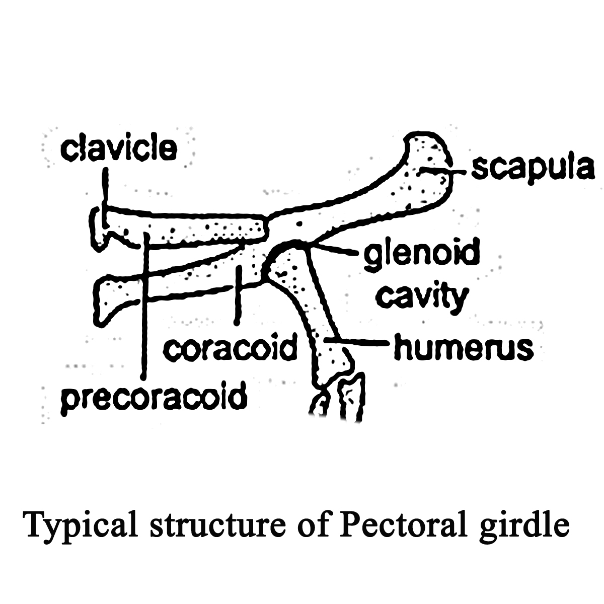
1. Scapula
2. Suprascapula
3. Coracoid
4. Clavicle
Scapula: In elasmobranchs, the two halve of pectoral girdles are cartilaginous, which are fused forming scapo-coracoid.
In bony fishes or teleosts, cleithrm is the basic unit of pectoral fins which supports the supracleithrum and through posttemporal attached directly to the skull.
In higher vertebrae, it is thin, flat and stout bone somewhat rectangular or triangular in shape. It lies on the dorsal the surface of the rib cage and helps to the attachment of forelimb with the body.
Supra-scapula : It is a thin, flat and cartilaginous structure attached distally to the Scapula. In the case of amphibian and reptiles, supra-scapula is rectangular or triangular in shape, covers thorax ventrally. In the case of Aves, the supra scapula is reduced or absent.
Coracoid: It is stout bone articulate with the scapula, to form a glenoid cavity. In glenoid cavity, the head of humor bone is articulate. The distal process of coracoid forms acrogoracoid process.
Clavicle: It is slender and elongated bone also called a color bone. It gives support to the pectoral girdle. In the case of the birds, clavicles unite to from forcula. In the carnivore, clavicle is reduced or lost.
Pelvic girdle:
It is also called as hip bone. It gives support to the hind limb and balances the weight of the body. The degree of development of pelvic girdle depends upon, whether the animal is a bipedal or quadruped.
Pelvic girdle consists of two equal halve. Each half is called as ossinnominatum. Each osinnominatum typically consists of the following parts.


a) Upper Ilium
b) Inner Ischium
c) Pubis
Ilium :
It is the elongated stout upper bone that, articulate with ischium and pubis to form acetabular cavity. In the acetabular cavity, the head of the femur bone is articulate. The ilium bone articulates with the sacral vertebra of the vertebral column.
In case of the frog, the ilium is elongated shows the presence of iliac crest. In reptiles and Aves, it is stout and elongate bone without any crest. In mammals, ilium is expanded fan like.
Ischium: It is the posterior-lateral segment of pelvic girdle. It has distal tuberosity called ischiatic tuberosity. To the ischiatic tuberosity muscle of thigh are attached.
Pubis. The pubis is the anterior portion of the hip / pelvic girdle. The two half of the pelvic girdles are attached by a pubic ligament (Reptile) or pubic symphysis in mammals. The ilium, ischium and pubis shows presence of large foramen called as obturator foramen through which nerve and blood vessels pass.
In case of fishes, the pelvic girdle gives support to the pelvic fins. In male inner edge of pelvic fin shows the presence of clasper which acts as a copulatory organ and helpful for sexual dimorphism.


No comments:
Post a Comment